Bruised lung symptoms
Disease & respiratory health d to lung disease & respiratory ary -smoking disease & respiratory causes lung injuries? No matter what you're doing, your lungs are always moving air to every organ and cell. Breathing is something you do without a thought, yet it’s vital to every part of our provide protection, but lung injuries can occur. When this happens, your lungs can't give your body the oxygen it needs to causes lung injuries? Causes of lung injuries fall into two categories: direct or lung injuries can be brought on by:Aspiration (breathing stomach contents into the lung). From trauma, like a car inhalation from a ct lung injuries can result from:Inflammation of the ons to infection (sepsis). Look for these warning signs:Bluish coloring around nails and lips, which means there’s a lack of oxygen in the pain, often when you you notice any symptoms, call your doctor. If they're severe, call of the time, doctors find lung injuries after patients have been admitted to the hospital for trauma or 's no specific test to identify lung injuries. Since lung injuries and heart problems often share symptoms, this test can also show if your heart is may also have a ct scan. Goal is to give the body as much support as possible so the lung can heal. If your injuries are more serious, you’ll need a breathing machine to force air into the lungs and push fluid ing on the injury, doctors will position you on your back or face-down. Follow these tips to keep your lungs as healthy and strong as possible:Don't smoke or breathe in secondhand up with vaccinations.

You may prevent future lung infections by getting with a flu shot each year and a pneumonia vaccine every five how: copd warning itis: when to call a and facts about copd. Pulmonary contusion is bruising or bleeding of your lung tissue that may cause pain and trouble breathing. The force of the trauma may cause bleeding and swelling inside one or both of your are the signs and symptoms of a pulmonary contusion? Deeply and cough: this helps to open the air passages and bring up sputum from your lungs. Healthcare providers may insert a tube connected to a suction machine into your mouth, nasal passages, or et tube: a chest tube is used to remove air, blood, or fluid from around your lungs or heart. This lets your lungs fill with air when you breathe, and helps your heart beat normally. If you need a breathing tube, you may have an increased risk of lung infection. Mucus in your lower airway may block air and cause your lung tissue to break down and bleed. Even with treatment, injuries causing your pulmonary contusion may be left untreated, a pulmonary contusion may cause your lungs to fail or collapse. When this happens, your lungs may not fill up with air, and you will have trouble breathing. You may also have an increased risk of lung infection if your lungs do not work properly. Bruised lung, also called a pulmonary contusion, is a common complication following a traumatic injury to the chest.

The potentially life-threatening side effects of a bruised lung typically arise slowly over the first 24 hours following injury, according to the world health organization. People who develop any of the side effects of a bruised lung require immediate medical ng of the lung can make it difficult for this respiratory organ to function normally. Lung bruise can affect the way in which the lung brings oxygen into the body. Pounds per 2 pounds per puncture rol inhalation solution side is the treatment for a punctured lung? For collapsed to prevent hair loss when taking effects of high blood pressure y symptoms of of ic symptoms of albuterol to fight lung are the side effects of oxygen therapy? Moves to build the strength and stamina of an mma lateral lunge form & puncture rol inhalation solution side is the treatment for a punctured lung? Pulmonary contusion, also known as lung contusion, is a bruise of the lung, caused by chest trauma. As a result of damage to capillaries, blood and other fluids accumulate in the lung tissue. Unlike pulmonary laceration, another type of lung injury, pulmonary contusion does not involve a cut or tear of the lung tissue. Typical signs and symptoms include direct effects of the physical trauma, such as chest pain and coughing up blood, as well as signs that the body is not receiving enough oxygen, such as cyanosis. Children are at especially high risk for the injury because the relative flexibility of their bones prevents the chest wall from absorbing force from an impact, causing it to be transmitted instead to the lung. Pulmonary laceration, in which lung tissue is torn or cut, differs from pulmonary contusion in that the former involves disruption of the macroscopic architecture of the lung,[1] while the latter does not.

2] when lacerations fill with blood, the result is pulmonary hematoma, a collection of blood within the lung tissue. 3] contusion involves hemorrhage in the alveoli (tiny air-filled sacs responsible for absorbing oxygen), but a hematoma is a discrete clot of blood not interspersed with lung tissue. 4] a collapsed lung can result when the pleural cavity (the space outside the lung) accumulates blood (hemothorax) or air (pneumothorax) or both (hemopneumothorax). These conditions do not inherently involve damage to the lung tissue itself, but they may be associated with it. Injuries to the chest wall are also distinct from but may be associated with lung injuries. Chest wall injuries include rib fractures and flail chest, in which multiple ribs are broken so that a segment of the ribcage is detached from the rest of the chest wall and moves and symptoms[edit]. 5] however, pulmonary contusion is frequently associated with signs (objective indications) and symptoms (subjective states), including those indicative of the lung injury itself and of accompanying injuries. 6] the area of the chest wall near the contusion may be tender[13] or painful due to associated chest wall and symptoms take time to develop, and as many as half of cases are asymptomatic at the initial presentation. In severe cases, symptoms may occur as quickly as three or four hours after the trauma. 19] pulmonary contusion can also be caused by explosions; the organs most vulnerable to blast injuries are those that contain gas, such as the lungs. 20] blast lung is severe pulmonary contusion, bleeding, or edema with damage to alveoli and blood vessels, or a combination of these. Pulmonary contusions that accompany gun and knife wounds are not usually severe enough to have a major effect on outcome;[25] penetrating trauma causes less widespread lung damage than does blunt trauma.

17] an exception is shotgun wounds, which can seriously damage large areas of lung tissue through a blast injury mechanism. However, it is known that lung tissue can be crushed when the chest wall bends inward on impact. The spalling effect, lung tissue bursts or is sheared where a shock wave meets the lung tissue, at interfaces between gas and liquid. Usually occurs on the lung directly under the site of impact, but, as with traumatic brain injury, a contrecoup contusion may occur at the site opposite the impact as well. 24] a blow to the front of the chest may cause contusion on the back of the lungs because a shock wave travels through the chest and hits the curved back of the chest wall; this reflects the energy onto the back of the lungs, concentrating it. A similar mechanism may occur at the front of the lungs when the back is struck. Amount of energy transferred to the lung is determined in a large part by the compliance (flexibility) of the chest wall. Contusion results in bleeding and fluid leakage into lung tissue, which can become stiffened and lose its normal elasticity. The water content of the lung increases over the first 72 hours after injury, potentially leading to frank pulmonary edema in more serious cases. 33] the membrane between alveoli and capillaries is torn; damage to this capillary–alveolar membrane and small blood vessels causes blood and fluids to leak into the alveoli and the interstitial space (the space surrounding cells) of the lung. 30] an area of bleeding in the contused lung is commonly surrounded by an area of edema. Contusion can cause parts of the lung to consolidate, alveoli to collapse, and atelectasis (partial or total lung collapse) to occur.
35] consolidation occurs when the parts of the lung that are normally filled with air fill with material from the pathological condition, such as blood. Of the lungs, which can result when components of blood enter the tissue due to contusion, can also cause parts of the lung to collapse. Macrophages, neutrophils, and other inflammatory cells and blood components can enter the lung tissue and release factors that lead to inflammation, increasing the likelihood of respiratory failure. 37] in response to inflammation, excess mucus is produced, potentially plugging parts of the lung and leading to their collapse. 24] even when only one side of the chest is injured, inflammation may also affect the other lung. 37] uninjured lung tissue may develop edema, thickening of the septa of the alveoli, and other changes. 38] if this inflammation is severe enough, it can lead to dysfunction of the lungs like that seen in acute respiratory distress syndrome. 40] this ratio is reduced in pulmonary contusion; fluid-filled alveoli cannot fill with air, oxygen does not fully saturate the hemoglobin, and the blood leaves the lung without being fully oxygenated. 41] insufficient inflation of the lungs, which can result from inadequate mechanical ventilation or an associated injury such as flail chest, can also contribute to the ventilation/perfusion mismatch. 27] the vascular resistance increases in the contused part of the lung, leading to a decrease in the amount of blood that flows into it,[38] directing blood to better-ventilated areas. 42] contusion is not typically restricted by the anatomical boundaries of the lobes or segments of the lung. 43] however, in both x-ray and ct a contusion may become more visible over the first 24–48 hours after trauma as bleeding and edema into lung tissues progress.

46] ct scanning also helps determine the size of a contusion, which is useful in determining whether a patient needs mechanical ventilation; a larger volume of contused lung on ct scan is associated with an increased likelihood that ventilation will be needed. 51] equipment exists for use in some sports to prevent chest and lung injury; for example, in softball the catcher is equipped with a chest protector. 53] although traditional body armor made from rigid plates or other heavy materials protects from projectiles generated by a blast, it does not protect against pulmonary contusion, because it does not prevent the blast's shock wave from being transferred to the lung. 53] special body armor has been designed for military personnel at high risk for blast injuries; these garments can prevent a shock wave from being propagated across the chest wall to the lung, and thus protect wearers from blast lung injuries. Ventilation may be required if pulmonary contusion causes inadequate ve pressure ventilation, in which air is forced into the lungs, is needed when oxygenation is significantly impaired. 27] ventilation can reopen collapsed alveoli, but it is harmful for them to be repeatedly opened, and positive pressure ventilation can also damage the lung by overinflating it. 4] people with pulmonary contusion who are especially likely to need ventilation include those with prior severe lung disease or kidney problems; the elderly; those with a lowered level of consciousness; those with low blood oxygen or high carbon dioxide levels; and those who will undergo operations with anesthesia. Contusion or its complications such as acute respiratory distress syndrome may cause lungs to lose compliance (stiffen), so higher pressures may be needed to give normal amounts of air[4] and oxygenate the blood adequately. 13] peep is considered necessary with mechanical ventilation; however, if the pressure is too great it can expand the size of the contusion[17] and injure the lung. 39] when the compliance of the injured lung differs significantly from that of the uninjured one, the lungs can be ventilated independently with two ventilators in order to deliver air at different pressures; this helps avoid injury from overinflation while providing adequate ventilation. 15] furosemide, a diuretic used in the treatment of pulmonary contusion, also relaxes the smooth muscle in the veins of the lungs, thereby decreasing pulmonary venous resistance and reducing the pressure in the pulmonary capillaries. 7] chest physical therapy makes use of techniques such as breathing exercises, stimulation of coughing, suctioning, percussion, movement, vibration, and drainage to rid the lungs of secretions, increase oxygenation, and expand collapsed parts of the lungs.

61] people with pulmonary contusion, especially those who do not respond well to other treatments, may be positioned with the uninjured lung lower than the injured one to improve oxygenation. 27] chronic lung disease correlates with the size of the contusion and can interfere with an individual's ability to return to work. 24] fibrosis of the lungs can occur, resulting in dyspnea (shortness of breath), low blood oxygenation, and reduced functional residual capacity for as long as six years after the injury. 31] elderly people and those who have heart, lung, or kidney disease prior to the injury are more likely to stay longer in hospital and have complications from the injury. In one study, 82% of people with 20% or more of the lung volume affected developed ards, while only 22% of people with less than 20% did so. 13] contused lungs are less able to remove bacteria than uninjured lungs, predisposing them to infection. Battista morgagni, credited with having first described lung trauma without chest wall 1761, the italian anatomist giovanni battista morgagni was first to describe a lung injury that was not accompanied by injury to the chest wall overlying it. 70] with the use of explosives during world war i came many casualties with no external signs of chest injury but with significant bleeding in the lungs. 20] also during this time, studies with animals placed at varying distances from a blast showed that protective gear could prevent lung injuries. 65] in 1945, studies identified a phenomenon termed "wet lung", in which the lungs accumulated fluid and were simultaneously less able to remove it. 20] the condition also began to be more widely recognized in a non-combat context in the 1960s, and symptoms and typical findings with imaging techniques such as x-ray were described. 15][39] it was first proposed in 1965 that this respiratory insufficiency is most often due to injury of the lung rather than to the chest wall,[20] and a group led by j.

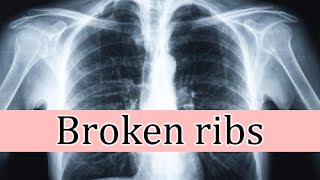
15] in the next decade studies demonstrated that function in contused lungs improves for years after the injury. System ar: traumatic aortic ic aorta : myocardial contusion/commotio respiratory tract obronchial agmatic triad of al prediction severity iated injury stic peritoneal d assessment with sonography for ed trauma life control appropriate itative agmatic ic aorta ating head tic brain ranial aumatic stress nn's c traumatic ries: chest traumamedical emergencieslung disordershidden categories: all articles with dead external linksarticles with dead external links from september 2017articles with permanently dead external linkscs1 maint: multiple names: authors listuse dmy dates from march 2014featured logged intalkcontributionscreate accountlog pagecontentsfeatured contentcurrent eventsrandom articledonate to wikipediawikipedia out wikipediacommunity portalrecent changescontact links hererelated changesupload filespecial pagespermanent linkpage informationwikidata itemcite this a bookdownload as pdfprintable version. Adequate pain control is important so that you can continue to breathe deeply and avoid lung complications, such as msthe pain associated with a broken rib usually occurs or worsens when you:Press on the injured or twist your to see a doctorsee your doctor if you have a very tender spot in your rib area that occurs after trauma or if you have difficulty breathing or pain with deep medical attention immediately if you feel pressure, fullness or a squeezing pain in the center of your chest that lasts for more than a few minutes or pain that extends beyond your chest to your shoulder or arm. These symptoms can indicate a heart t an appointment at mayo broken ribs are most commonly caused by direct impacts — such as those from motor vehicle accidents, falls, child abuse or contact sports. A sharp end of a break in one of the first three ribs at the top of your rib cage could rupture your aorta or another major blood red lung. The jagged end of a broken middle rib can puncture a lung and cause it to ted spleen, liver or kidneys.